Technology has led to the creation of improvements and innovations that have permanently altered the way dental professionals deliver patient care.
We strive to continually provide our patients with the highest level of service and expertise to which they have become accustomed, the following digital technologies are now part of our daily operations.
Digital dental technologies enable consultations with patients and timely collaborations with other dental specialties. This allows improved quality of care, enhanced diagnosis, and more precision in treatment planning.
CBCT Scan
Cone Beam Computed Tomography Scan
Commonly referred to as a CT or CAT scan. The CT images provide a detailed three- dimensional view of the head, neck, and the oral regions to be examined.

A CBCT scan is prescribed to identify critical anatomic landmarks and structures such as:
- tooth root fracture or resorption
- tooth root position
- bone density
- Temporo-Mandibular Joint (TMJ) anatomy and health
- Jawbone pathology
- Jaw nerve location
- Sinus floor location, shape, and pathology
Additionally, the 3-dimensional (CBCT) images are used for Computer Guided Implant Surgery (CGIS) planning. CGIS is the virtual planning for precise dental implant placement. This helps to ensure optimal long term functional and esthetic outcomes.
CBCT is painless, noninvasive, and accurate, it provides more information than conventional digital x-rays and can image bone & soft tissue.
The CBCT scanner also emits less radiation than other traditional medical grade CT scans or x-rays.
Digital Intra-oral Scanner
The digital intra-oral scanner provides a new, comfortable, and precise way to take impressions of patients' teeth.

Impressions have historically involved making a mold of a patient's mouth using trays filled with a soft, messy, and often bad-tasting material that would harden around patients' teeth and gums. The digital impression eliminates the “gooey” mess and gag-reflex often encountered. It's just more comfortable!
The digital models are exact replicas of a patient's mouth used to plan precision surgical and restorative treatments.
In addition to being much more comfortable for the patient, using the scanner allows us to be more efficient, saving the patient additional visits.
Digital X-Rays
The improved diagnostic capability of traditional digital dental x-rays is well known and a “standard of care” today. The ability to view and manipulate x-rays on a large screen allows the patient to better visualize and understand their treatment plan.
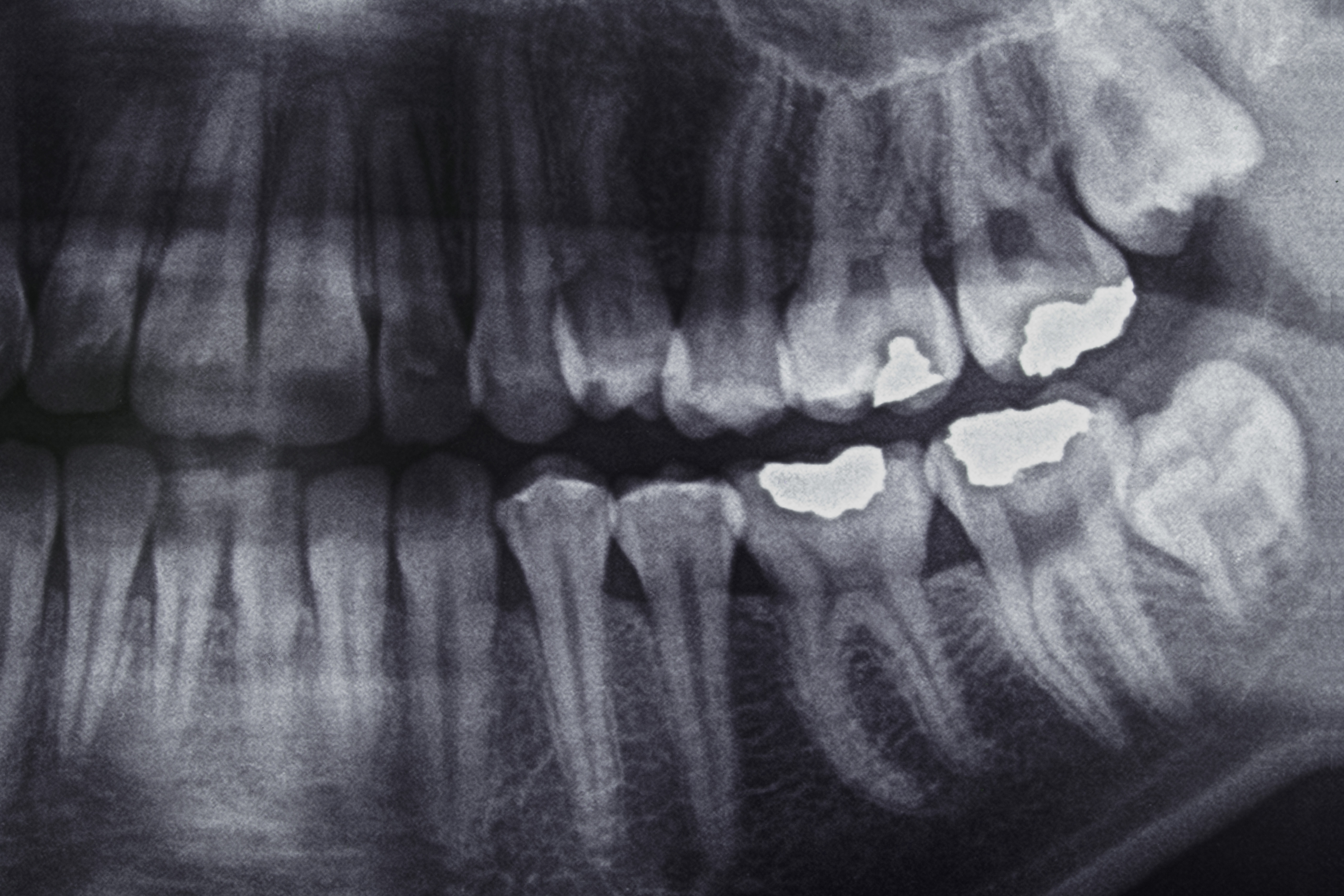
Digital x-rays are instant and no longer require chemicals to develop the film. The elimination of film and chemical disposal contribute to a cleaner environment.
Digital dental x rays also reduce the amount of radiation exposure (90% below traditional film x-rays).
Platelet Rich Fibrin (PRF)
Platelet Rich Fibrin is a healing biomaterial with great potential for bone and soft tissue regeneration.

The PRF slowly releases growth factor proteins to assist in tissue healing .and regeneration without inflammation. Dr Franzetti uses this process in combination with bone grafts, and soft tissue surgical procedures promoting hemostasis and bone growth.
A minimal amount of patient's own blood is drawn and spun in a centrifuge to extract high concentrations of platelets which contain protein growth factors. These proteins are essential to the healing process of soft tissue and bone. PRF aids patients to heal from dental procedures more quickly.


